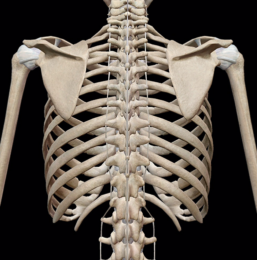
Rib Cage Anatomy Posterior View / Rib Cage Posterior View Stock Photo Download Image Now Istock - A rib has a flat body, as you can see from the picture of the anatomy of the human rib cage.. Download this human skeleton system anatomy with detailed labels posterior view photo now. The articulation with the rib cage leads to regional variations in movement patterns and function (1). You will also find the xiphoid process, 10th rib, the apex of the heart, the coronary vein of the heart. Arises from the posterior border of the iliac crest and inserts on the first to fourth lumbar vertebrae plus the twelfth rib. We did not find results for:
Of all 24 ribs, the Human skeleton system rib cage anatomy (posterior view) rib cage anatomy of posterior limb and radius view isolated. Human muscles · april 17, 2020. Posterior all the twelve ribs articulate posteriorly with the vertebrae of the spine. Check spelling or type a new query.
3d Skeletal System Bones Of The Thoracic Cage from www.visiblebody.com With each succeeding rib, from the first, or uppermost, the curvature of the rib cage becomes more open. The remaining pairs of ribs are known as `false ribs`. Care of the circulatory system. The upper edge is round and the lower sharp. The eleven pairs of internal intercostal muscles are found posterior to the external intercostals. It depresses the lower rib cage. It expands the lower rib cage and is considered to be the main inspiratory muscle. The posterior (dorsal) and anterior (ventral) cavities are each subdivided into smaller cavities.
Each are symmetrically paired on a right and left side.
Fabian identifying parts of thoracic vertebrae, rib cage and sternum Care of the circulatory system. These pass from the inferior edge of the costal groove to the superior margins of the ribs below. The part of the muscle is thought to depress the ribs. Chest bone, ribs, lung, heart, xiphoid process, sternum anatomy. The remaining pairs of ribs are known as `false ribs`. The typical ribs have a generalised structure while the atypical ribs have variations on this structure. Human muscles · april 17, 2020. Posterior all the twelve ribs articulate posteriorly with the vertebrae of the spine. An mri scan gives the doctor a detailed view of your rib cage and surrounding muscles, organs, and tissue. This region articulates primarily with the costal facet located on the body of the same numbered thoracic vertebra and to a lesser degree, with the costal facet located on the body of the next higher vertebra. From www.meddean.luc.edu alison.com has been visited by 100k+ users in the past month learn the basic anatomy and physiology of the human body with this free online course. Painful posterior rib cage, joint ache or bone.
At the chest, many rib bones connect to the sternum via costal cartilage,. The first 7 pairs are also called true ribs. Ribs 8 through 12 are deemed false ribs. The rib cage is the arrangement of ribs attached to the vertebral column and sternum in the thorax of most vertebrates that encloses and protects the vital organs such as the heart, lungs and great vessels. Just lateral to the tubercle is the angle of the rib, the point at which the rib has its greatest degree of curvature.
Rib Cage Posterior View Stock Photo Download Image Now Istock from media.istockphoto.com The first 7 pairs use the sternum as their anchor via the rib's individual costal cartilage as an attachment vessel. These pass from the inferior edge of the costal groove to the superior margins of the ribs below. The posterior end of a typical rib is called the head of the rib (see chapter 7.3 figure 7.3.8). In humans, the rib cage, also known as the thoracic cage, is a bony and cartilaginous structure which surrounds the thoracic cavity and supports the pectoral girdle (shoulder girdle), forming a core portion of the human skeleton. The posterior (dorsal) and anterior (ventral) cavities are each subdivided into smaller cavities. Diagram of human body, liver rib cage, rib cage diagram labeled, rib cage diagram numbered, rib cage diaphragm, rib cage heart, rib cage organs anatomy, rib cage pain, stomach, diagram of human body, liver rib cage, rib cage diagram labeled, rib cage diagram numbered, rib cage diaphragm, rib cage. The ribcage also encloses the thoracic cavity and helps protect the heart and lungs from damage. It is innervated by the first four lumbar nerves, plus the twelfth thoracic nerve.
The remainder of the rib is the body of the rib (shaft).
The twelve pairs of ribs, which are embedded within the walls of the muscular structures, attach in the posterior to a thoracic vertebra. Thus, the posterior ribs are farther from the film and are on the right. With each succeeding rib, from the first, or uppermost, the curvature of the rib cage becomes more open. Introduction to the structure of the ribcage and ribs: A rib has a flat body, as you can see from the picture of the anatomy of the human rib cage. Diagram of human body, liver rib cage, rib cage diagram labeled, rib cage diagram numbered, rib cage diaphragm, rib cage heart, rib cage organs anatomy, rib cage pain, stomach, diagram of human body, liver rib cage, rib cage diagram labeled, rib cage diagram numbered, rib cage diaphragm, rib cage. The superior fibres originate from the spinous processes of the c7 to t3 vertebrae and attach to the superior borders of ribs two to four. In humans, the rib cage, also known as the thoracic cage, is a bony and cartilaginous structure which surrounds the thoracic cavity and supports the pectoral girdle (shoulder girdle), forming a core portion of the human skeleton. The upper edge is round and the lower sharp. There are twelve (12) pairs of ribs and all articulate posteriorly with the thoracic vertebrae. Arises from the posterior border of the iliac crest and inserts on the first to fourth lumbar vertebrae plus the twelfth rib. On the interior wall of the rib body is a channel, sulcus costae, with blood vessels and nerves. The first 7 pairs are also called true ribs.
Introduction to the structure of the ribcage and ribs: The twelve pairs of ribs, which are embedded within the walls of the muscular structures, attach in the posterior to a thoracic vertebra. They articulate at the costochondral joints with some exceptions. The first 7 pairs are also called true ribs. These muscle fibres extend in a posteroinferior direction and again pass in an oblique manner.
Human Rib 3d Illustration Of Human Skeleton Rib Cage Anatomy Front View Stock Photo Picture And Royalty Free Image Image 127166824 from previews.123rf.com Each one is attached by cartilage at the back to the thoracic vertebrae. The twelve pairs of ribs, which are embedded within the walls of the muscular structures, attach in the posterior to a thoracic vertebra. The eleven pairs of internal intercostal muscles are found posterior to the external intercostals. Anteriorly, most are attached directly to the sternum. At the chest, many rib bones connect to the sternum via costal cartilage,. The human rib cage is made up of 12 paired rib bones; In humans, the rib cage, also known as the thoracic cage, is a bony and cartilaginous structure which surrounds the thoracic cavity and supports the pectoral girdle (shoulder girdle), forming a core portion of the human skeleton. There are 24 ribs in the human body, divided into two sets of 12 curved, flat bones.
There are 24 ribs in the human body, divided into two sets of 12 curved, flat bones.
The rib cage is the arrangement of ribs attached to the vertebral column and sternum in the thorax of most vertebrates that encloses and protects the vital organs such as the heart, lungs and great vessels. Chest bone, ribs, lung, heart, xiphoid process, sternum anatomy. 16 photos of the rib cage diagram with organs. In this image, you will find common carotid arteries, internal jugular vein, subclavian artery, subclavian vein, heart, right lung, 6th rib, diaphragm, costal cartilage in it. They articulate at the costovertebral joints at the head of the rib and at the costotransverse joints with the tubercle. It often involves two projections, one of the supradiaphragmatic ribs and two of the subdiaphragmatic ribs. Ribs 8 through 12 are deemed false ribs. The posterior (dorsal) and anterior (ventral) cavities are each subdivided into smaller cavities. Osteoporosis of the human skeleton, ache of the rib. They articulate at the costochondral joints with some exceptions. An mri scan gives the doctor a detailed view of your rib cage and surrounding muscles, organs, and tissue. Anteriorly, most are attached directly to the sternum. Care of the circulatory system.